...
Overview

What is Fetal Echocardiography?

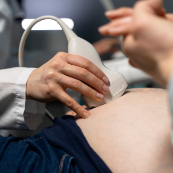
Fetal Echocardiography is a specialized ultrasound performed between 18 to 24 weeks of pregnancy to examine the baby’s heart structure, function, and blood flow. It is an essential screening tool to detect congenital heart defects (CHDs) and ensure optimal heart health before birth.

Why Choose

Why Choose Racham for Fetal Echocardiography?

At Racham Fetal Medicine Centre, Bangalore, we provide.

Advanced Imaging Technology
Advanced Technology

High-resolution ultrasound for precise heart imaging.

Expert Fetal Medicine Specialists
Expert Fetal Specialists

Experienced doctors specializing in fetal cardiology.

Comprehensive Pregnancy Monitoring
Comprehensive Diagnostics

Detailed assessment of fetal heart structures and blood flow.

Trusted Care
Patient-Centric Approach

Compassionate, stress-free experience for expectant parents.

Condition

Findings of Fetal Echocardiography

Fetal Echocardiography provides detailed insights into the baby’s heart development, including:

Detecting Structural Abnormalities
Fetal Development
Fetal Development

Congenital Heart Defects (CHDs) – Detecting Structural Abnormalities

This scan detects heart defects such as:

  • Ventricular Septal Defects (VSD) – Small or large holes in the heart.
  • Atrioventricular Septal Defects (AVSD) – Improperly formed heart chambers.
  • Hypoplastic Left Heart Syndrome (HLHS) – Underdeveloped left heart structures.
  • Tetralogy of Fallot (TOF) – A combination of four heart defects affecting blood flow.

Early detection helps in planning specialized care, potential interventions, and delivery strategies for better outcomes.

Ensuring Proper Cardiac Function

Heart Rhythm Abnormalities – Ensuring Proper Cardiac Function

Fetal Echocardiography detects arrhythmias (irregular heartbeats) such as:

  • Tachycardia – Faster-than-normal heartbeat.
  • Bradycardia – Slower-than-normal heartbeat.
  • Heart block – Disruptions in electrical signals controlling the heartbeat.

Identifying rhythm abnormalities early allows for timely medical management to improve fetal health.

Blood Flow & Valve Function
Amniotic Fluid Disorders

Blood Flow & Valve Function – Evaluating Circulatory Efficiency

This scan assesses:

  • Aortic and Pulmonary Valve Function – Ensuring proper blood flow.
  • Doppler Flow Analysis – Evaluating oxygen and nutrient supply to the baby.
  • Coarctation of the Aorta (CoA) – Narrowing of the major artery, which may require post-birth intervention.

Timely diagnosis allows parents and doctors to plan the best course of action for delivery and treatment.

Fetal Aortic Doppler
Heartbeat Detection
Heartbeat Detection

Fetal Cardiac Tumors – Detecting Rare Abnormalities

Some rare tumors, such as rhabdomyomas, can develop in the baby’s heart. This scan helps identify such conditions and aids in prenatal monitoring and postnatal treatment planning.


Why is Fetal Echocardiography Important?

Fetal Echocardiography in Bangalore helps expectant parents understand their baby’s heart health. Key benefits include:

  • Early Detection of Heart Defects – Enables timely planning of care and interventions.
  • Assessment of Fetal Circulation – Ensures normal blood flow and heart function.
  • Monitoring High-Risk Pregnancies – Essential for mothers with diabetes, lupus, or a history of CHDs.
  • Guidance for Delivery Planning – Helps decide if specialized medical support is needed at birth.

Who Should Get a Fetal Echocardiography?

This scan is recommended for pregnant women who:

  • Have a family history of congenital heart disease.
  • Are above 35 years of age (advanced maternal age).
  • Have diabetes, high blood pressure, or lupus.
  • Have had infections or medication exposure that could affect the baby’s heart.
  • Had abnormal findings in previous scans or screenings.

At Racham Fetal Medicine Centre, Bengaluru, our specialists guide expectant mothers with expert counseling and support.


racham-sidebar-image
Free Online Consultation With Fetal Medicine Experts
Book Appointment

Process

What Happens During a Fetal Echocardiography?

1

Preparation

No special preparation is required. A moderately full bladder may help.

Preparation

2

Scan Process

A transducer is moved over the abdomen to capture detailed heart images.

Real-Time Insights

3

Detailed Examination

Our specialists analyze heart structures, function, and blood flow.

The Scan

4

Results & Consultation

Findings are discussed immediately, and parents receive a detailed report.

Comprehensive Report

Videos

Expert Care for Fetal Health

Informative ultrasound insights on fetal heart, brain, and pregnancy scans.

  • Fetal Heart - Cardiac screening At anomaly scan
    Cardiac screening At anomaly scan

    Fetal heart view in anomaly scan.

  • Fetal Brain @ Anomaly scan
    Fetal Brain @ Anomaly scan

    Ultrasound to check fetal brain development.

  • Pregnancy ultrasound
    Pregnancy ultrasound

    Pregnancy ultrasound monitors fetal development

  • Fetal Echo - Tetralogy of Fallot
    Fetal Echo - Tetralogy of Fallot

    Fetal Echo detects Tetralogy of Fallot in the fetus.


What Our Patients Say

Real experiences from patients on our specialized care.

google Review us on Google
Book an Appointment

Schedule Your Consultation at Racham Fetal Medicine Centre

https://www.rachamfetalmedicine.com/wp-content/uploads/2024/11/racham-fetal.png
https://www.google.com/maps/embed?pb=!1m18!1m12!1m3!1d497698.7749125013!2d77.30125893383429!3d12.95445953963007!2m3!1f0!2f0!3f0!3m2!1i1024!2i768!4f13.1!3m3!1m2!1s0x3bae1670c9b44e6d%3A0xf8dfc3e8517e4fe0!2sBengaluru%2C%20Karnataka!5e0!3m2!1sen!2sin!4v1732880805311!5m2!1sen!2sin

    FAQ's

    Frequently Asked Questions – Fetal Echocardiography

    Fetal Echocardiography is an advanced ultrasound that evaluates the baby’s heart structure, function, and blood flow between 18-24 weeks of pregnancy.

    It helps detect congenital heart defects and rhythm abnormalities early, allowing for better management and treatment planning before birth.

    No, it is a non-invasive, painless ultrasound procedure that uses high-frequency sound waves.

    The scan typically takes 30-45 minutes, depending on fetal position and scan complexity.

    Yes, fetal echocardiography is a completely safe ultrasound with no known risks to the baby.

    While highly accurate, some conditions may develop later in pregnancy or after birth, requiring further evaluation.

    Our specialists will discuss the findings, recommend further tests if needed, and guide you on the best prenatal and postnatal care options.

    The cost may vary depending on the clinic and ultrasound type. At Racham Fetal Medicine Centre, we offer affordable, high-quality scans. Contact us for pricing details.

    NO. Gender determination is illegal in India under the PCPNDT Act. Kindly do not request gender disclosure.

    To schedule your Fetal Echocardiography at Racham Fetal Medicine Centre, call us at +918095950077 or email rachamfetalmedicine@gmail.com. Our team will guide you through the process and answer all your queries.
    Home About Contact Inquiry
    Seraphinite AcceleratorOptimized by Seraphinite Accelerator
    Turns on site high speed to be attractive for people and search engines.