- Anjali Verma
Dr. Dolly Chavda provided exceptional care during my pregnancy. Her expertise in fetal medicine gave me peace of mind, and the entire team was incredibly supportive.

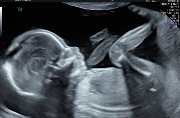
The Nuchal Translucency (NT) Scan, also known as the First Trimester Screening (FTS), is a specialized ultrasound performed between 11 to 13+6 weeks of pregnancy. This scan measures the fluid at the back of the baby’s neck (nuchal translucency) to assess the risk of chromosomal abnormalities like Down syndrome (Trisomy 21), Edward’s syndrome (Trisomy 18), and Patau’s syndrome (Trisomy 13). It also evaluates early fetal development, detecting major structural anomalies and providing crucial insights into placental health through uterine artery Doppler screening to predict preeclampsia risk.

At Racham Fetal Medicine Centre, Bengaluru, we provide:

High-resolution imaging for precise NT measurement and fetal assessment.

Experienced fetal medicine specialists ensuring accurate interpretation.
NT Scan combined with Double Marker Test for enhanced risk assessment.
Years of reliable, precise, and ethical prenatal screening in Bangalore.
High-resolution imaging for precise NT measurement and fetal assessment.
Experienced fetal medicine specialists ensuring accurate interpretation.
NT Scan combined with Double Marker Test for enhanced risk assessment.
Years of reliable, precise, and ethical prenatal screening in Bangalore.
The NT Scan in Bangalore provides vital early pregnancy insights, including:


The NT Scan evaluates the thickness of fluid at the back of the baby’s neck. Increased NT thickness may indicate a higher risk of Down syndrome (Trisomy 21), Trisomy 18, or Trisomy 13.
The NT Scan also helps detect major structural defects such as:

The NT Scan includes uterine artery Doppler assessment to evaluate placental blood flow and predict the risk of preeclampsia, fetal growth restriction (FGR), and preterm birth. Early detection allows for preventive measures to ensure a healthy pregnancy.
The NT Scan in Bengaluru provides critical early pregnancy insights, helping expectant parents:
During the NT Scan in Bangalore, our specialists assess:
The NT Scan in Bangalore is recommended for all pregnant women, particularly those:

At Racham Fetal Medicine Centre, Bengaluru, the NT Scan is a painless and non-invasive ultrasound:
No special preparation is needed, but a Partially filled bladder may help.

A transducer is gently moved over the abdomen to capture fetal images.

NT thickness and fetal structures are evaluated.

The findings are explained immediately, with further recommendations if needed.

Informative ultrasound insights on fetal heart, brain, and pregnancy scans.

Fetal heart view in anomaly scan.

Ultrasound to check fetal brain development.

Pregnancy ultrasound monitors fetal development

Fetal Echo detects Tetralogy of Fallot in the fetus.
Visual insights into expert prenatal care and patient trust.


Dr. Dolly Chavda provided exceptional care during my pregnancy. Her expertise in fetal medicine gave me peace of mind, and the entire team was incredibly supportive.
I’m grateful for the accurate diagnosis and treatment we received at Racham Fetal Medicine. Dr. Chavda’s professionalism and compassion were outstanding throughout the process.
Thanks to Dr. Dolly, my high-risk pregnancy was managed flawlessly. The scans and advice were precise, and I felt confident every step of the way.
We were impressed with the advanced diagnostics and the way the staff explained everything step by step. Thank you for making us feel so cared for!

Here are some commonly asked questions about the Viability Scan to help you understand the process and benefits of this crucial early-stage pregnancy test:
 Optimized by Seraphinite Accelerator